What is the purpose of the joint ultrasound examination?
A joint ultrasound examination is a special ultrasound, which painlessly maps the soft tissues, muscles, tendons and their adhesion surrounding the joints (shoulders, elbows, wrists, hips, knees, ankles), the cartilage covering the bone and the surrounding skeletal muscles.
The examination also reveals inflammation around the joints, injuries and rupture in the muscles and tendons, thickening of the synovial membranes, calcifications that may be present, and bleeding and fluid build-up.
Its advantage over X-ray and CT scans is that it does not involve radiation exposure and the joints can be examined while moving. However, since ultrasound is not suitable for the examination of bony patterns, for the detection of possible fractures or deformities, it is recommended in all cases in addition to X-ray, CT or MRI examination.
Which lesions can be examined with ultrasound?
Joint ultrasound is recommended if a physical examination or X-ray or CT scan does not give a clear answer to the cause of the joint complaints. Joint ultrasound can help diagnose the following lesions:

- determination of injuries: ligament injuries, muscle injuries, haematomas can be clearly identified
- degenerative joint diseases: the thinning of the cartilage covering the bones becomes visible on ultrasound, as well as the narrowing of the articular gap
- identification of inflammatory joint diseases: thickening of the synovial membrane, the amount of fluid build-up, and erosion on the surface of the bones all indicate inflammation.
- diagnosis of intra-articular nerve injuries: it helps diagnose carpal tunnel syndromes
- Since the surrounding formulas are also examined during the ultrasound examination, it is visible during the examination if the pain is transmitted following inflammation of one of the surrounding organs.
- It is also suitable for post-operative follow-up, such as after a knee replacement.
How is the joint ultrasound examination performed?
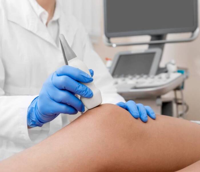
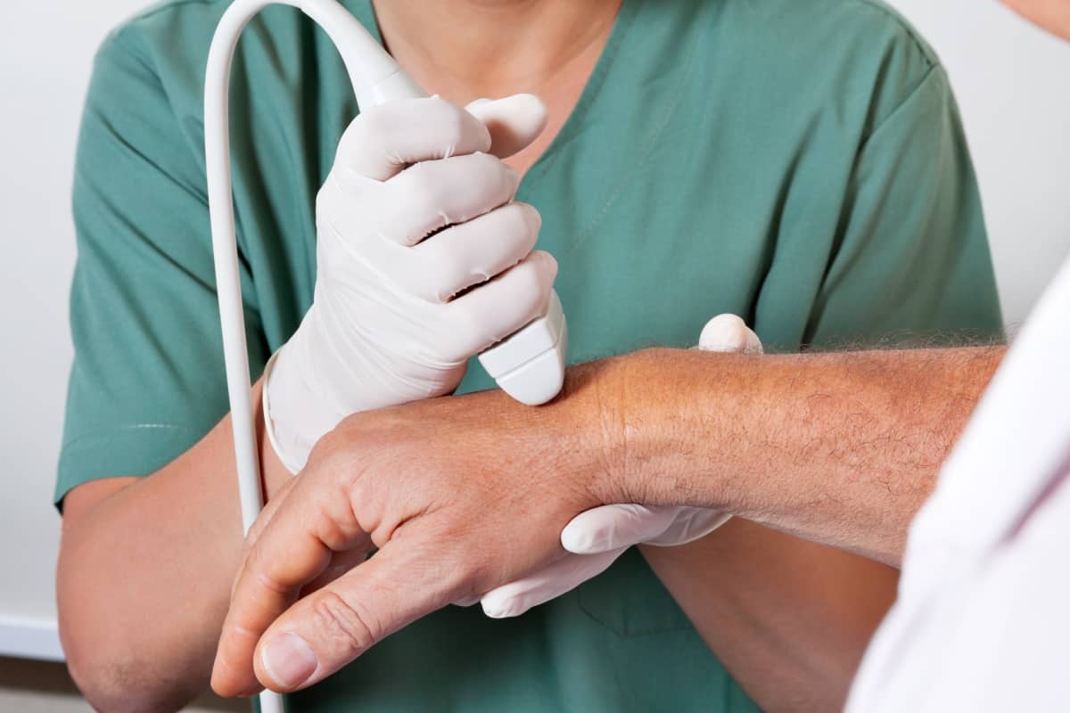
The examination is performed in a sitting or lying position, depending on the location of the joint to be examined. To perform the examination, the area of the joint must be exposed, after which the examining doctor will apply a gel to the skin to help the probe function properly. The specialist will review the joint and surrounding formulas, which will take about 10-15 minutes.
The total duration of the examination is approx. 20 minutes.
How to prepare for the joint ultrasound?

The examination does not require special preparation on the part of the patient, but it is advisable to arrive in clothing that can be easily removed from the body part to be examined.
