Breast tomosynthesis
In Hungary, breast cancer is the most common cancer among women, which means an increased health risk for women over the age of 40.
The sooner a breast tumour is recognized, the more effective the treatment is. That is why it is extremely important that in addition to the annual, regular gynaecological screening, women also participate in regular breast screening.
Two methods can be used to screen for breast cancer. Without complaints, conventional two-dimensional mammography is recommended. In case of a complaint or any lesion, three-dimensional breast tomosynthesis is recommended, which is currently the most modern and reliable diagnostic test.
What is the difference between breast tomosynthesis and mammography?
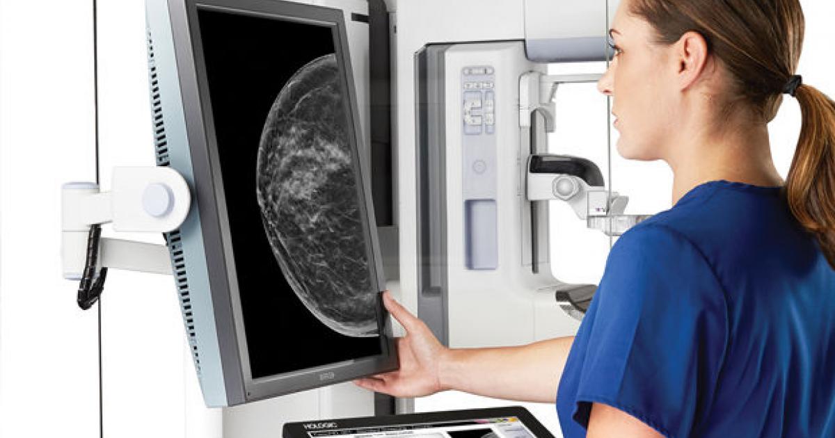
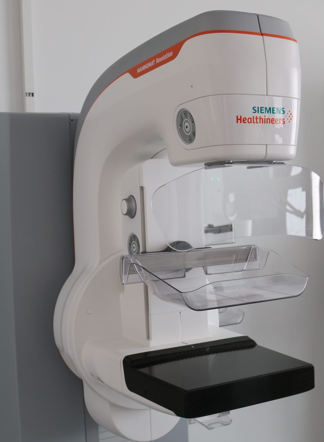
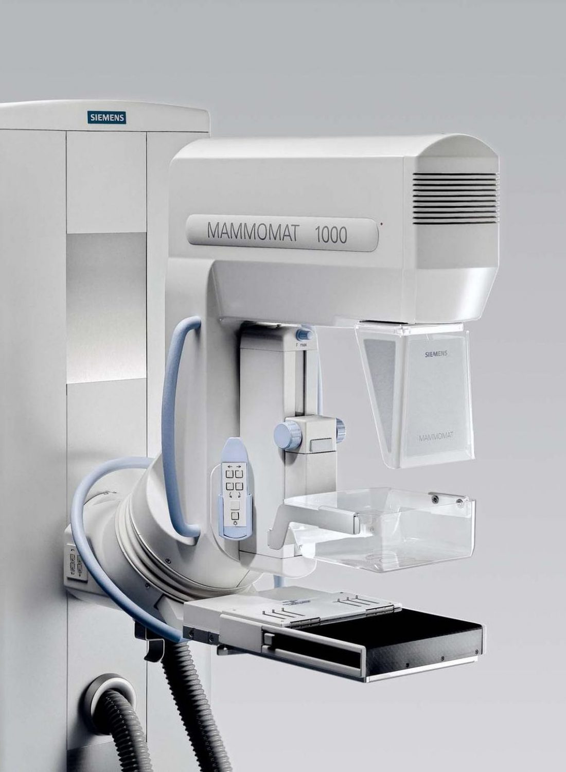
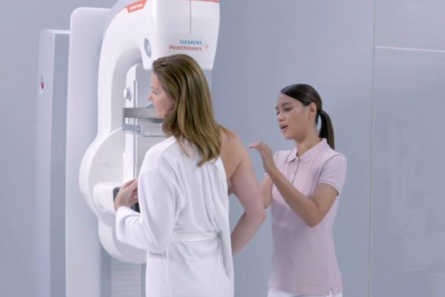
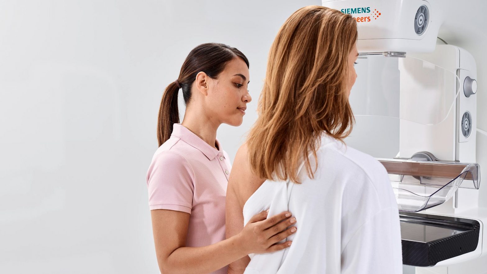
The Medicover Diagnostic Center features state-of-the-art, three-dimensional breast tomosynthesis equipment.
Breast tomosynthesis compared to conventional mammography:
- can detect 41.5% more invasive types of breast cancers
- is less uncomfortable due to milder breast compression
- takes less time
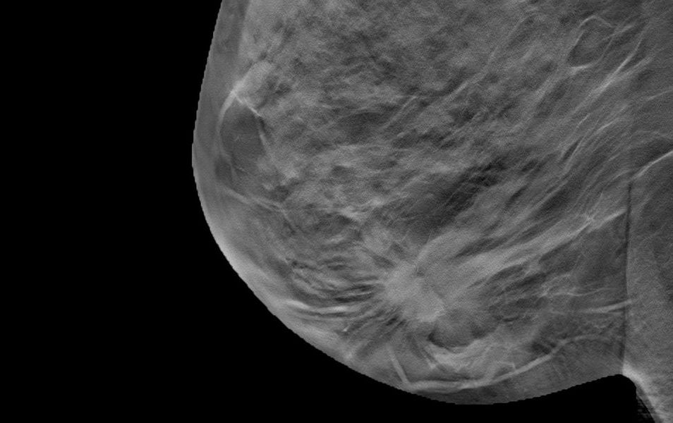
- provides the highest resolution and most detailed image available in three dimensions
- collects data in the widest 50-degree angle range currently available, from which it uses computer software to take up to 15 images per breast with a slice thickness of 1 millimetre (the number of images depends on the size of the breasts) compared to 2 images taken during conventional mammography.
| TOMOSYNTHESIS | MAMMOGRAPHY | |
| Breast tomosynthesis can detect 41.5% more invasive types of breast cancer than mammography. | ||
| 3D | Imaging | 2D |
| 50° | Test angle range | – |
| personalized compression | Chest pressure | unpleasant feeling of compression |
| yes | Tissue sampling possibility | yes |
| yes | Automatic breast tissue density measurement | no |
How is a breast tomosynthesis examination performed?
The examination does not require any prior preparation, cosmetics that have come into contact with the skin must be wiped off beforehand.
For the examination of the breasts, the upper body must be exposed and then you have to stand in front of the examination device in accordance with the instructions of the radiology assistant, after which the breasts are compressed and examined.
No further action is required after the examination.
When is it recommended to have the tomosynthesis performed?
The examination should be performed in the first half of the cycle, i.e. within two weeks of the first day of menstruation. In case of a breast implant, the tomosynthesis is much more effective than conventional mammography.
Can the examination be performed while breastfeeding?
The examination can also be performed when it comes to breast-feeding mothers, but breast-feeding should not be resumed until 24 hours after the examination.
When is the result expected?
The completed recording is reviewed by a breast specialist radiologist. The result is available in our online system 3-4 working days after the test.
What are the benefits of breast tomosynthesis?
Minor chest compression
Most women are deterred from regular breast screening by the severe and unpleasant pain they experience during a common mammography. During breast tomosynthesis, thanks to the so-called personalized tender compression, the pressure on the breasts is lighter and automatically adjusted to their size, making the examination less inconvenient than mammography.
The best quality and highest resolution image available
In contrast to the traditional two-dimensional mammography examination, which only takes a two-sided image of the breasts, breast tomosynthesis provides the best quality and most detailed image available in Hungary.
It collects data in the widest 50-degree angle range currently available, from which it uses computer software to take up to 15 images per breast with a slice thickness of 1 millimetre (the number of images depends on the size of the breasts) compared to 2 images taken during common mammography.
Tissue sampling (stereotaxic breast biopsy)
With current mammographic procedures available in most locations, samples taken during a biopsy are analysed by another device or a special tissue sample scanner, usually in another room, after the examination.
This is accompanied by a longer waiting time and an uncomfortable posture during the examination.
Based on the evaluation of the results of the tomosynthesis examination, the radiologist may, if appropriate, recommend tissue sampling.
The great advantage of breast tomosynthesis over common mammography is that the analysis of the samples can be started up to 20 seconds after the tissue sampling – the device indicates whether the tissue sample is suitable for histological analysis, i.e. whether it contains the altered tissue of the sample – thus reducing examination and waiting times.
Advantages of tomosynthesis-guided stereotaxic sampling:
The biggest advantage of breast tomosynthesis is its ability to detect the early stages of breast cancer that may develop. The procedure can be used to determine exactly where the microcalcification is, even the size of a pinhead, inside the breast tissue.
These very tiny calcium deposits are not visible in common mammographic images or are indistinguishable from healthy breast tissues. Malignancies can develop from microcalcifications in the breasts.
Tomosynthesis-guided stereotaxic sampling is the only diagnostic procedure available that allows histological determination of microcalcification even without surgery.
Automatic breast tissue density measurement
Women with denser breast tissue are at higher risk of developing breast cancer. Furthermore, it is also more likely that the tumour is difficult to identify due to dense breast tissue.
Determining breast tissue density is thus one of the most important part of the breast cancer diagnosis process.
In case of common mammography examinations, the radiologist tries to estimate the density of breast tissue by analysing the image. The breast tomosynthesis device at Medicover Diagnostic Center is able to automatically determine breast tissue density during the examination.