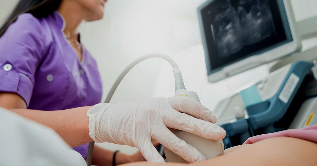
Pregnancy ultrasound examination
The primary goal of a pregnancy ultrasound examination is to screen for foetal abnormalities as soon as possible and with the greatest possible accuracy, as well as to shed light on the health status of the mother.
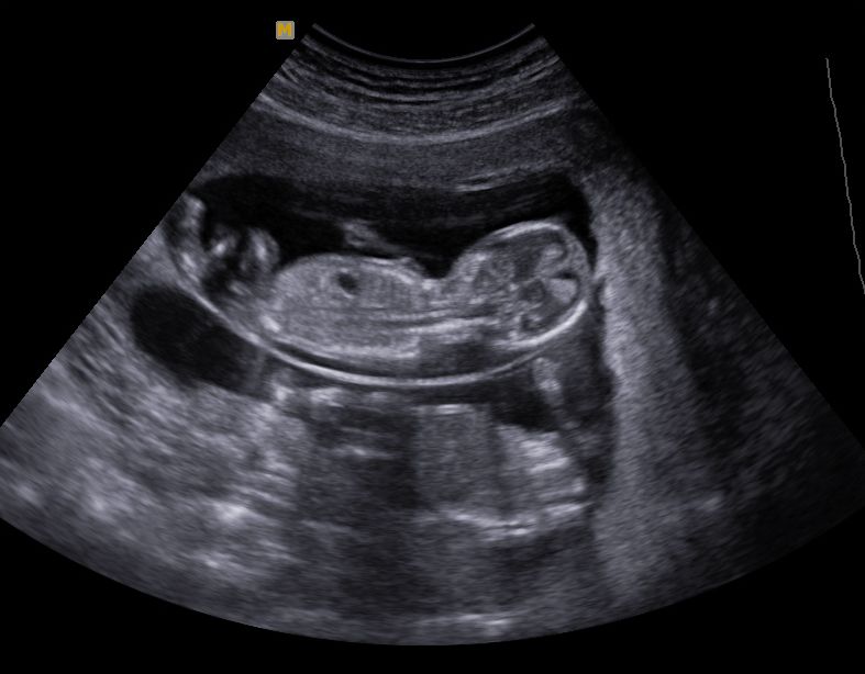
Proof of pregnancy can be provided at the earliest in the second week after the absence of menstrual bleeding, if a pregnancy test performed at home has shown a positive result. During regular imaging examinations, we can monitor the maturity of the foetus, changes in its size, its location within the uterus, we can determine its sex over time, and we can also screen for any developmental abnormalities.
Which areas does a pregnancy ultrasound examine?
The pregnancy ultrasound (which can take the form of both vaginal and abdominal ultrasound) examines the health status of all pelvic organs (uterus, ovaries, and fallopian tubes).
Vaginal ultrasound is used to diagnose lesions in the pelvis, and at the same time, abdominal ultrasound can be used to detect abnormalities outside the pelvis.
Both examinations are completely painless, do not involve radiation exposure, and have no foetal harm.
How is a pregnancy ultrasound performed?
- The first ultrasound examination is performed vaginally to determine pregnancy between its 5-10 weeks. The aim of the examination is to confirm the pregnancy within the uterus, to determine the number of foetuses, and to determine the exact time of pregnancy.
- The second examination takes place in week 12-13 of pregnancy, to reveal signs of developmental and chromosomal abnormalities.
- The third examination is performed at the 18th week of pregnancy, which is performed through the abdominal wall. In addition to the examination of the foetal skull, chest, heart, abdominal organs, spine and limbs, the amount of placenta and amniotic fluid is also determined.
Please be sure to bring the questionnaire that you have completed during the ultrasound examination of weeks 12-18!
- The fourth ultrasound examination is performed at week 30. The aim is to monitor the proper development of the foetus and to screen any abnormalities that may be detected later.
- The last examination is performed in the 36th week of pregnancy, the aim of which is to screen the birth risk group for the purpose of planning the delivery: to determine the position and expected weight of the foetus in uterus, and to examine the placenta and amniotic fluid.
What can we learn from a pregnancy ultrasound examination?

- the size of the foetus,
- the weight of the foetus,
- the age of the foetus can be determined,
- developmental disorders can be screened,
- abnormalities in the mother’s body can also be screened,
- the location and condition of the placenta can be determined,
- twin pregnancies can be detected,
- the location of the foetus and placenta can be accurately detected before the amniotic fluid is examined.
How to prepare for a pregnancy ultrasound?
The examination does not require special preparations. In the case of vaginal ultrasound, the emptied bladder, and in the case of abdominal ultrasound, the full bladder, helps the specialist performing the ultrasound examination. However, prior to the examination, it is advisable to clarify in advance with the doctor ordering the examination whether you should arrive with a full or empty bladder.
