Lesser pelvis MRI examination
A lesser pelvis MRI scan gives a detailed picture of the internal genitals (ovaries, uterus, prostate), bladder and rectum.
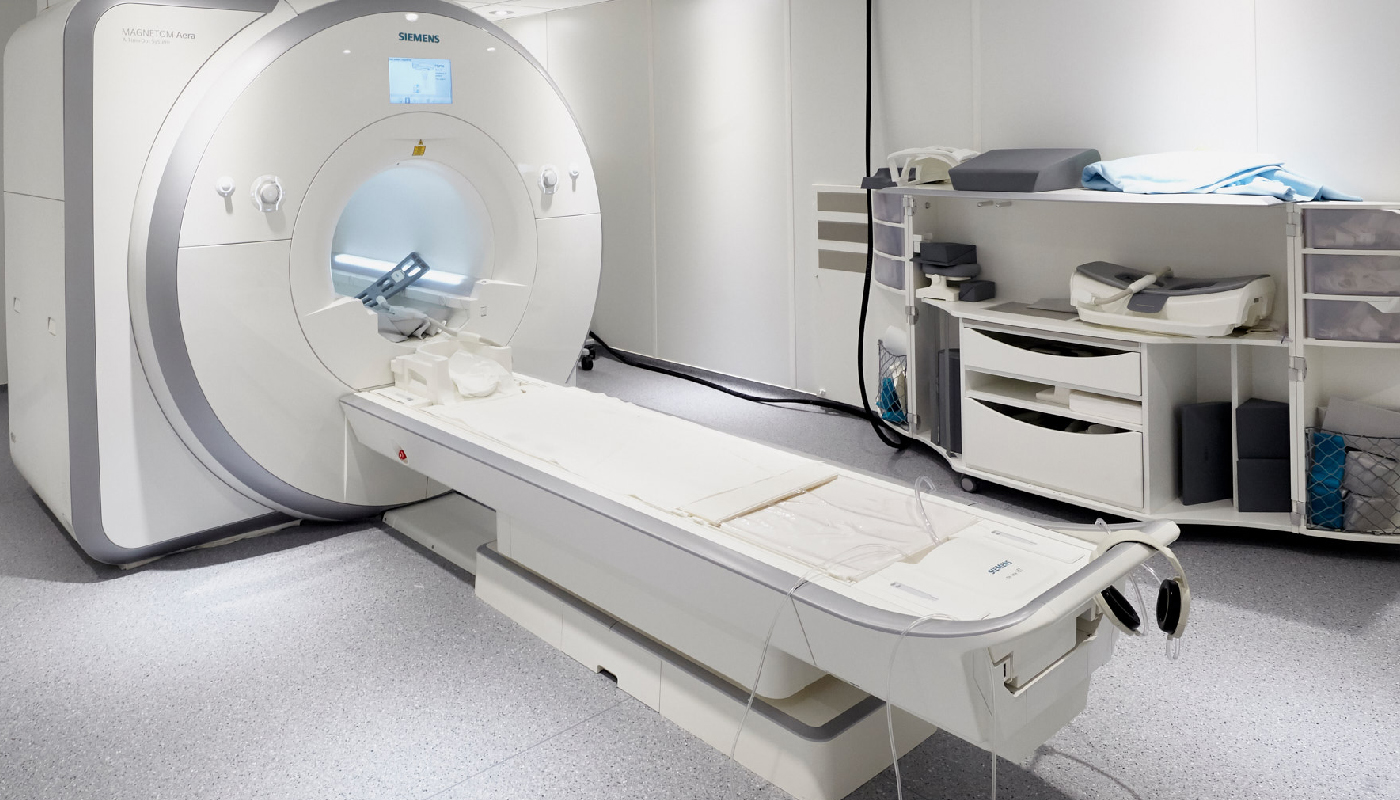
MRI is an imaging diagnostic procedure, which uses magnetic field to take detailed images of the human body.
The magnetic field generated by the device converts the energy changes in the body into a detailed, three-dimensional image by a high-performance computer.
What types of diseases does the examination help diagnose?
A lesser pelvis MRI is usually prescribed by a specialist to detect gynaecological or urological diseases:
- kidney disease (kidney cyst, suspected tumour)
- urinary problems (incontinence)
- adrenal disease
- bladder lesions
- gynaecological diseases: lesions of the ovaries and uterus, endometriosis
- in men, the cause of prostate enlargement
In case of which symptoms is it recommended to have the scan done?
A gynaecologist may request an examination in addition to laboratory tests and ultrasound if the following complaints exist:
- in case of lower abdominal and pelvic pain, and cramps of unknown origin
- painful menstruation
- bleeding disorders
- infertility
A urologist usually recommends an examination for the following complaints:
- decreased urine output, nocturnal urination
- difficult, erratic urination
- urinary incontinence
- abdominal pain
- low back pain
How to prepare for the examination?
In all cases, it is recommended to consult a gynaecologist or urologist before a lesser pelvis MRI examination, depending on the complaints.
A moderately full bladder is optimal for the examination, which in women also helps the uterus to have adequate visibility on the images.
Due to the strong magnetic field, it is forbidden to bring any metal, device or object containing metal into the examination room! This also applies to implants in the body, as well as to jewellery and piercings.
Medical recommendation is always required for a scan with contrast agent. Do not eat for 6 hours before the examination.
The condition for the administration of the contrast agent is the appropriate kidney function value (creatinine and urea). This requires a recent laboratory finding, but it is also possible to perform a rapid creatinine test and eGFR measurement on site with the help of an MRI operator, which is done from the fingertip with a prick.
How is the examination performed?
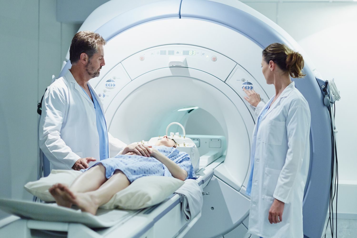
The lesser pelvis MRI scan is performed in supine position.
The operator will place the required coil and then the examination bed will lift you into the magnetic tube.
It is important that you remain still during certain parts of the examination, you can only change your posture with the permission of the operator. During the scan, you can communicate with our colleague continuously via the built-in microphone and speaker, or you can interrupt it by pressing the panic button.
The MRI device is noisy, a knocking, clicking, and machine sound can be heard during the examination, the intensity of which can be reduced by using earplugs.
The test lasts for 50 minutes.
When can the examination not be performed?
In case of non-MRI compatible metal, implant, prosthesis (pacemaker, orthopaedic metal: screw, plate, nail, wire), implanted joint prosthesis, prosthetic limb, implanted defibrillator, neurostimulator, implanted drug dispenser, vascular implants (e.g. clips, vascular occlusion devices), ear implants, metal foreign body (e.g. bullet, splinter, other electronic implants) the test cannot be performed. These can move out of place during the test in a strong magnetic field and cause serious injury.
Therefore, in case of an implant, implanted metal, prosthesis, please bring a written certificate of its compatibility with MRI examination (certified by the implanting specialist, institution) for the examination itself, which clearly states: the suitability to enter the MRI examination room, to perform the MRI examination and marking the maximum magnetic field strength (1.5 Tesla, 3 Tesla) for the device or implant. Inform the person performing the examination about the implant beforehand.
In case of a scan with contrast agent, the examination is not performed with poor kidney function.
In case of pregnancy, the scan cannot be performed in the first trimester.
Due to the fixed diameter of the closed tube, it is not possible to perform the examination above 200 kg or depending on the abdominal circumference.
What can I expect after the examination?
The lesser pelvis MRI scan has no side effects.
The contrast agent used for MRI examinations is gadolinium-based. In rare cases, hypersensitivity might happen, which may occur in the form of an allergic reaction within 20 minutes after the administration of the contrast agent.
When is the result expected?
The completed recording is reviewed by the radiologist and the result will be available in our online system after 5 working days.
In all cases, consult the gynaecologist or urologist who ordered the examination to make a diagnosis and determine the appropriate treatment.