Mammography
Breast cancer is the most frequent tumorous disease among women aged 45-65 years.
Its development could be easily prevented by participating in regular screenings because if cancer is diagnosed in time, it has a good chance of being cured.
Self-examination plays an important role in prevention, as well as additional examinations such as breast ultrasound and mammography (common mammography, breast tomosynthesis).
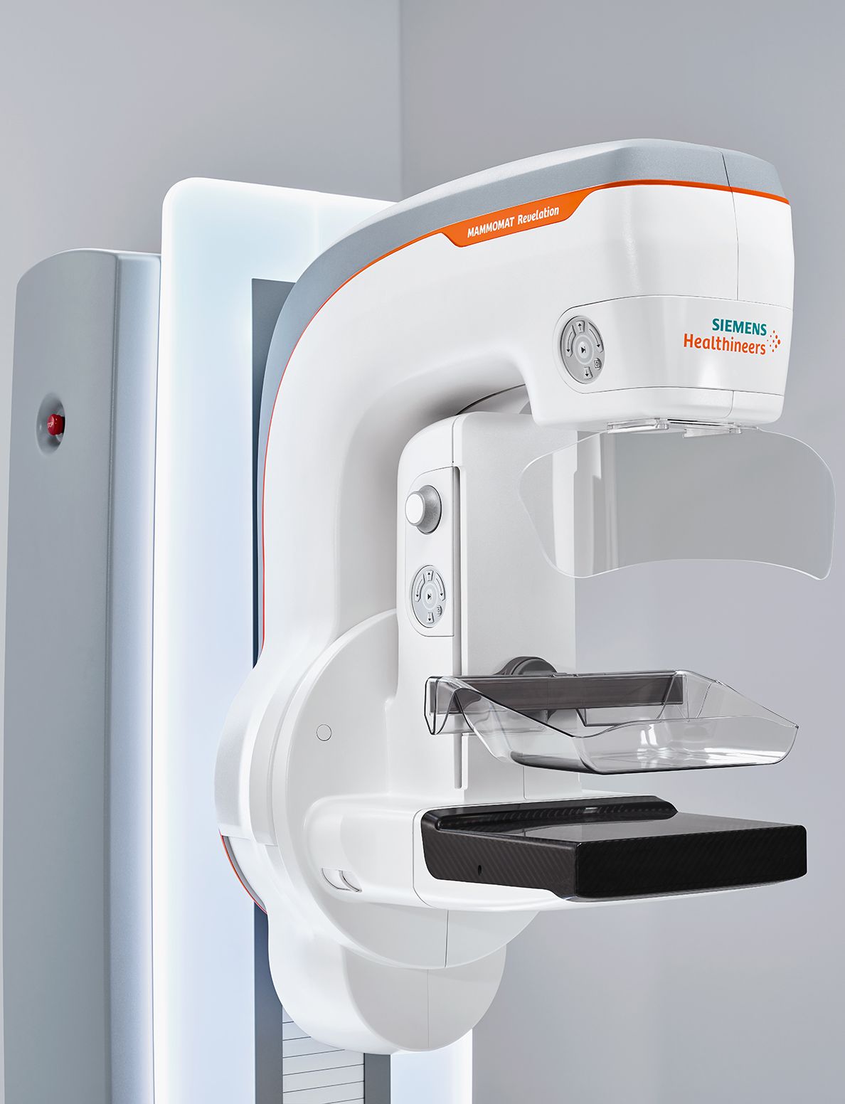
What is the difference between breast tomosynthesis and common mammography?
The Medicover Diagnostic Center features state-of-the-art, three-dimensional breast tomosynthesis equipment that is capable of detecting 41.5% more invasive types of breast cancer than traditional mammography.
| TOMOSYNTHESIS | MAMMOGRAPHY | |
| Breast tomosynthesis can detect 41.5% more invasive types of breast cancer than mammography. | ||
| 3D | Imaging | 2D |
| 50° | Test angle range | – |
| personalized compression | Chest pressure | unpleasant feeling of compression |
| yes | Tissue sampling possibility | yes |
| yes | Automatic breast tissue density measurement | no |
Advantages of breast tomosynthesis over common mammography
Breast tomosynthesis compared to common mammography:
- can detect 41.5% more invasive types of breast cancer
- is less uncomfortable due to milder breast compression
- takes less time
- provides the highest resolution and most detailed image available in three dimensions
- collects data in the widest 50-degree angle range currently available, from which it uses computer software to take a maximum of 15 images per breast with a slice thickness of 1 millimetre (the number of images depends on the size of the breasts) compared to 2 images taken during common mammography.
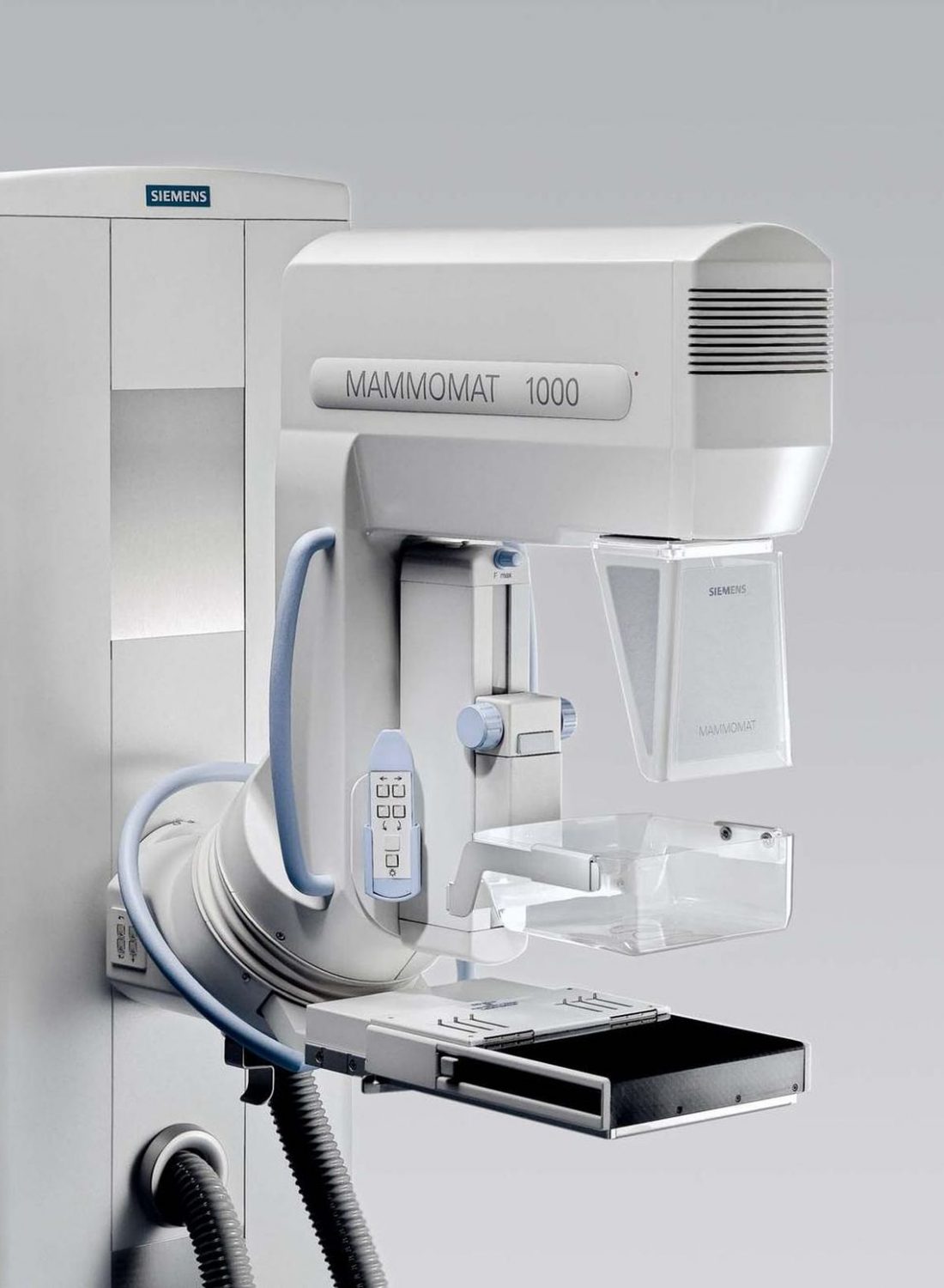
Common mammography
During screening, two-way mammography is performed on both breasts, preceded by physical examination, individual and family history, and risk factor assessment. It takes about 5 minutes to take a mammography.
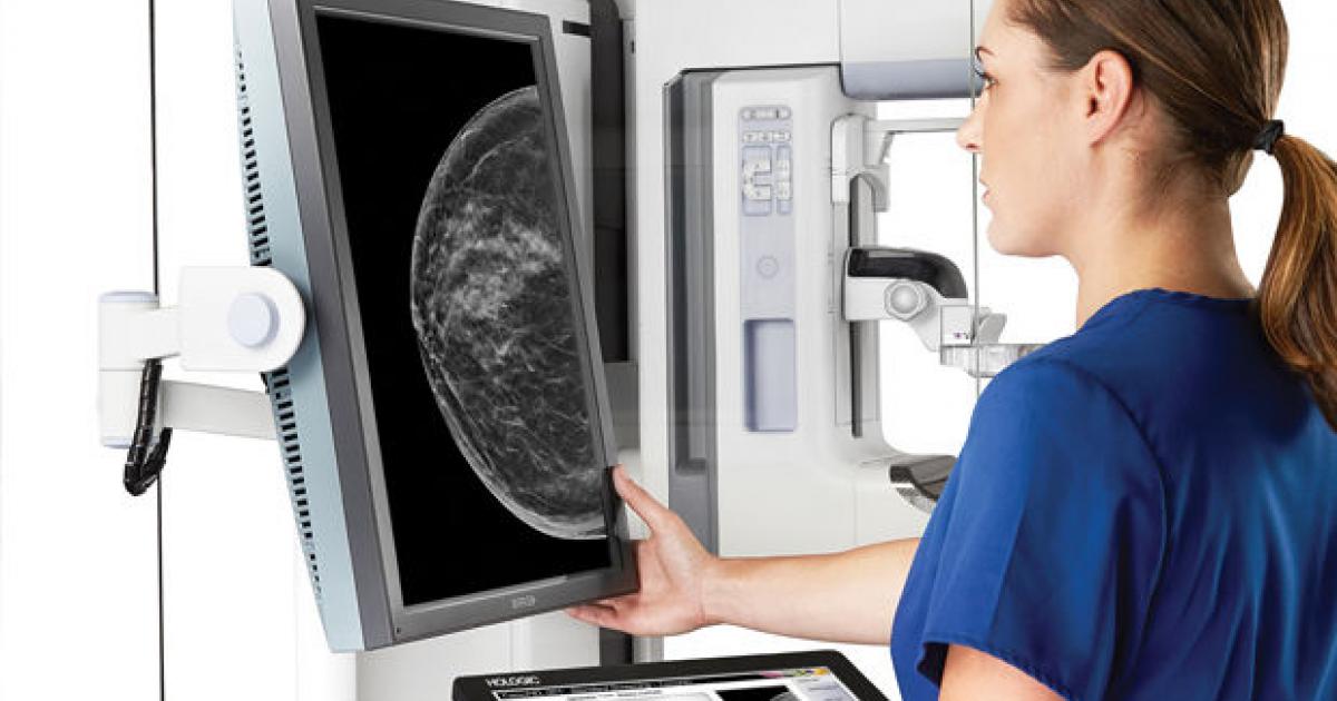
How does the examination go?
The upper body should be exposed so that the breasts can be examined. Then stand in front of the X-ray machine and place the breasts on the surface containing the X-ray film. The imaging process is performed with low-energy ionizing radiation, so the radiation exposure is small. The recordings are then evaluated by a specialist. The results are available in our online system 3-4 working days after the examination.
In some cases, an additional examination is needed to make an accurate diagnosis, which may be an additional special X-ray or ultrasound examination.
How can I have a common mammography?
The examination requires a paper-based medical referral, which is prescribed by your GP or a specialist. With aging, the risk of developing breast cancer increases. Women between the ages of 45 and 65 have the highest risk factor, so they need to be screened at least every two years. Of course, whatever suspicion arises about the presence of breast cancer, it is possible to perform the test regardless of age.
Common mammography or breast tomosynthesis?
Breast tomosynthesis is a more advanced method of examining breasts than common mammography. The image taken during tomosynthesis has a much higher resolution and is more detailed than the one taken during a common mammographic examination, which allows the early detection of breast lesions. In case of a tomosynthesis, breast compression is also much milder, making it less uncomfortable for the patient.