Paediatric radiology
Ultrasound is one of the radiological tests that is painless, does not involve radiation exposure, can be repeated an unlimited number of times, and can be safely performed in children. It is excellent for screening or diagnostic purposes in the event of complaints. It is mainly used to examine soft tissues, abdominal organs, and joints.
When might the examination be necessary?
The ultrasound scan can be requested by the parent as a screening test, and at 6-8 weeks of age, an ultrasound scan of the skull, abdomen, and hips is recommended.
It may be indicated by a specialist as part of a more complex examination, for example as part of a gastroenterological, endocrinological, or neurological examination.
It can also be performed in the event of complaints: either in case of acute, severe abdominal pain lasting several weeks, possibly accompanied by fever and bloody stools, or in case of acute or chronic joint pain, or to diagnose abnormalities or protrusions growing on the body.
How to prepare for the examination?
Usually, no special preparation is needed for an ultrasound scan.
It is not necessary to come to the examination on an empty stomach, but 1-2 hours before the abdominal ultrasound examination the child should drink only liquids. Avoid fizzy drinks on the day of the examination. The gall bladder and bile ducts can be optimally examined after 4-6 hours of fasting. For the examination of pelvic organs, urinary tract, and bladder, children should arrive with a full bladder and drink plenty of fluids half an hour before the examination, as the pelvic organs can be examined only to a limited extent if the bladder is empty.
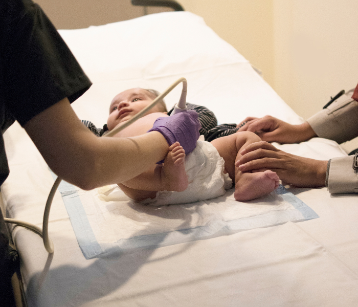
For an infant screening, it is preferable if the baby eats before the screening – it is easier to examine a satisfied, full and calm baby.
You are welcome to bring a pacifier, toys and music for the babies and children!
How is the examination carried out?
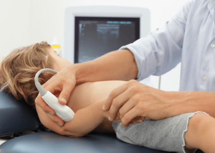
The scan is painless, and the ultrasound probe is used to scan the area to be examined using an ultrasound gel. Most of the scans are done in a lying position, and young children may find that their toys, music, and pacifiers help them to cope with lying down in unfamiliar surroundings, which is why we ask that parents are present at all times during the scan. The examination itself may take around 5-20 minutes, depending on the body region and any abnormalities found.
What to do after the examination?
After the examination, if an abnormality is found, or if you have requested an ultrasound scan as part of a complex examination by another specialist or your GP, please contact your referring doctor or GP with the findings to discuss further action.
What happens during an infant ultrasound screening (skull + abdomen + hip)?
Optimally, screening ultrasound scans of the skull, abdomen and hips are recommended at 6-8 weeks of age. Infant ultrasound screening is used to detect various developmental abnormalities not detected in utero as early as possible. It can avoid or reduce the risk of future, potentially permanent damage by early intervention.
Ultrasound scan of the skull
A cranial ultrasound scan examines the baby’s brain, through the large cranial cavity in the top of the head. Ultrasound can be used to bring part of the brain into view, so that certain developmental abnormalities, the consequences of infections, or some of the bleeding inside the skull during birth, and other abnormalities such as cerebral ventricular dilatation can be detected and monitored. If abnormalities are found during the examination, in some cases further neurological and possibly additional imaging studies (MRI scan) are recommended, in other cases ultrasound follow-up is sufficient.
The cranial cavity gradually narrows as the child grows and closes between 12 and 18 months of age. Above 6 months of age, if the cranial cavity is narrowed, ultrasound of the skull is limited, but in some cases, it can provide additional information up to 12 months of age.
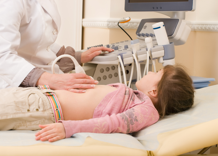
Abdominal ultrasound
During the screening abdominal ultrasound scan, we mainly examine the abdominal organs (liver, spleen, pancreas, kidneys, adrenal glands), as well as certain diseases and abnormalities of the intestines, stomach, and bladder. During the screening ultrasound scan, the abdominal organs are examined with the same thoroughness and detail as in an abdominal examination later in life.
Read more about abdominal ultrasound >>
Hip ultrasound
An ultrasound scan of the hip joint can help in the diagnosis of suspected congenital hip dislocation. From the age of 6 weeks, the so-called Graf angles are used to detect the abnormality and determine the severity of the deformity. The test should not be carried out in the first month of life, because at this age the newborn baby and its tissues and joints are still growing and maturing very rapidly. After the fourth month, the head of the femur starts to ossify, which makes ultrasound measurements difficult, so it is advisable to carry out the screening test before the fourth month, in order to allow for the earliest possible intervention.
Hip dislocation (hip dysplasia) is when the hip joint socket is “shallower” than normal, i.e. the socket does not wrap around the femoral head properly. The consequence of this is that the femoral head may move out of the joint more easily and more often (most often when the child is on its feet) and in cases of low dysplasia, the increased joint stress will lead to early arthrosis, a disease in which the joint wears out. Early therapy (starting in the second month of life) can eliminate or minimise these later complications.
What can be examined with abdominal ultrasound?
Liver and biliary tract: ultrasound scans are used to assess the size and structure of the liver, to look for so-called circumscribed abnormalities in the liver, such as cysts, vascular masses, benign or malignant tumours, and to examine the blood vessels supplying the liver. We then look for the gall bladder, gallstones, possible signs of inflammation and look at the bile ducts inside and outside the liver.
Ultrasound examination of the stomach: in children aged 2-8 weeks, the presence of projectile vomiting after repeated feedings should always be considered in the case of a condition known as pyloric stenosis. The muscles that regulate the emptying of the gastric outlet become thickened and obstruct the emptying of gastric contents. This results in frequent vomiting after feeding and consequent weight loss. Ultrasound examination of the sphincter allows rapid diagnosis of the condition.
Other diseases of the stomach cannot be investigated by ultrasound because of the air content.
Ultrasound is not suitable for the diagnosis of reflux disease!
Reflux is caused by the opening of the sphincter between the border of the oesophagus and the stomach, which causes stomach contents to back up into the oesophagus. In young babies, the same thing happens during a fall, which does not necessarily indicate an abnormal condition.
During an ultrasound scan, if we observe this area continuously for up to 1-2 minutes but do not see any reflux of stomach contents, we cannot rule out that this is not happening at other times, to an abnormal extent. Likewise, if we see a backflow, it does not mean that it is happening too often or that it is causing a complaint for the baby.
If reflux disease is suspected, a gastroenterology consultation is recommended.
Kidneys and urinary tract: the ultrasound scan shows the exact location, size and structure of the kidneys.
Kidney complaints in children may be due to kidney stones or, in the case of urinary tract infections, to inflammation of the renal pelvis. In the case of renal pelvis inflammation, ultrasound scans should be used to rule out or identify any predisposing developmental abnormalities or complications of the infection. Ultrasound can also be used to detect abnormalities such as dilatation of the renal pelvis and ureter, or certain developmental abnormalities. These include, for example, strictures in different parts of the urinary system or vesicoureteral reflux disease. This means that urine from the bladder flows back towards the kidneys.
High levels of dilatation and urinary reflux increase the pressure in the kidneys, which can lead to damage to the kidney tissue. It is therefore extremely important to detect urinary tract abnormalities early, because in the long term these conditions can lead to recurrent infections and damage to the kidneys.
Spleen: enlargement of the spleen can be a symptom of many conditions. In some infectious diseases (viral infections, especially mononucleosis, sepsis), haematopoietic disorders and other rare diseases based on genetic abnormalities, spleen enlargement may be seen on ultrasound examination in association with liver disease.
Intestine: ultrasound examination is an excellent way to examine the inflammation of the intestines. Active inflammation is indicated by the presence of thickening of the intestinal wall. Ultrasound is essential for the diagnosis and follow-up of severe intestinal infections or inflammatory bowel diseases (Crohn’s disease, ulcerative colitis).
In children, ultrasound is also the first choice for acute abdominal pathologies such as volvulus and invagination. Both conditions can cause intestinal necrosis through intestinal obstruction of the blood supply.
Appendicitis: this condition should be considered in all cases of abdominal pain, as it can have a wide variety of symptoms, none of which are typical. It is not in fact a disease of the coecum, but of the vermiform appendix, which is connected to it. An ultrasound scan may show a widened vermiform appendix as a sign of inflammation. It is often surrounded by inflammatory fluid and enlarged lymph nodes. Appendicitis can lead to peritonitis, abscesses, and sepsis, so early detection is very important. An ultrasound scan is essential for the diagnosis. It is important to stress, however, that the cecum, when placed behind another intestinal loop, cannot always be visualised by ultrasound.
Ultrasound examination of the pelvis
Uterus and ovaries: the investigation of hormonal abnormalities and blood disorders is now inconceivable without pelvic ultrasound. In the case of abdominal pains, in addition to appendicitis, gynaecological causes should always be considered as well, for example ovarian torsion. This condition also requires rapid diagnosis, as it can have serious consequences.