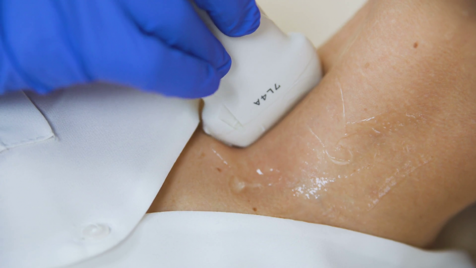
Cervical soft tissue ultrasound examination
During the ultrasound examination of the soft tissue of the neck, we examine the palpable nodules, superficial lesions and structural differences of the muscles on the neck and under the skin. Examination of the submandibular glands and parotid gland, sialolithiasis, possible inflammations, benign and malignant tumours can also be detected with the help of cervical ultrasound.
During the observation and examination of the salivary glands and cervical lymph nodes, we monitor their size, structure, vascularity, their lesions, and their relationship to their environment.

When is a cervical soft tissue ultrasound recommended?
Ultrasound examination of the soft tissue of the neck is most often used in the case of cervical, swallowing discomfort and painful (inflamed) salivary glands.
It is also used to monitor a known disease, as a follow-up examination for diagnosed cancer or surgery.
Cervical ultrasound is often performed during screening tests and as a health check.
Other ultrasound examinations of the neck:
Diseases, lesions and structural abnormalities of the thyroid gland are monitored by ultrasound examination of the thyroid gland. Vascular calcifications, the vessel stenosis caused by them, and the cervical narrowing caused by hypertension are performed with the ultrasound examination of the carotid arteries.
How to prepare for the cervical ultrasound?
The examination does not require special preparation. During the painless ultrasound examination, the patient lies on their back by placing a pillow under their shoulder in order for the neck to stretch well.
The examination is usually done without clothes on the upper body to protect it. A paper towel is placed on the shoulders as the ultrasound probe is lubricated with ultrasound gel to help the spreading the ultrasound waves.

