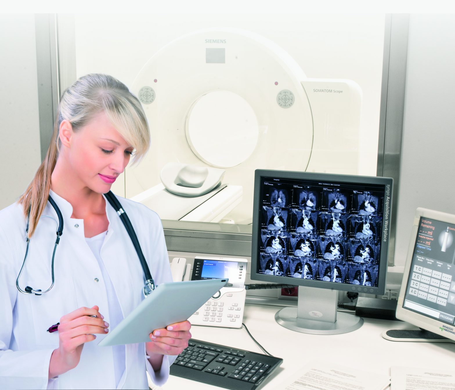
Abdominal and lesser pelvis CT scan
Abdominal and lesser pelvic CT can be used to examine lesions and diseases of all organs in the abdominal cavity and lesser pelvis. As diseases of the abdominal organs can be very diverse, it is recommended to consult an internal medicine specialist before a CT scan or to have an abdominal ultrasound examination. With these, it is much more effective to focus on a given area with a CT scan, as the more accurate the preliminary diagnosis, the more accurate your finding will be. If this is not possible and ultrasound recording is not available, the examination shall be performed according to the general professional protocol.
The CT device breaks down the body part to be examined into image slices a few millimetres in size, and then maps these slices by emitting x-rays. These x-rays pass through the body and are absorbed differently in each tissue and organ. The rays passing through the tissues are captured by the X-ray detector, and the signals thus obtained are converted into an image by the computer. With the help of the examination, cancerous and pathological lesions, bleeding, internal injuries, and inflammations can also be seen.
As the CT scan involves radiation, a specialist consultation is always required before the scan, where the clinician will consider whether the scan is necessary based on the symptoms. Due to radiation exposure, a CT scan should only be performed on the basis of a medical recommendation.
What types of diseases do abdominal and pelvic CT scans help diagnose?

Your doctor may ask you to have an abdominal and pelvic CT scan to diagnose the following conditions.
- Most cancers can only be safely analysed by CT, so the assessment of the possibility of surgery also relies on this method.
- search for metastases
- mapping of inflammatory processes
- mapping of bleeding
In case of which symptoms is it recommended to have the scan performed?
Your doctor may consider an examination if you have the potential to develop cancer, inflammation, bleeding, and your problem has not previously been confirmed with certainty with other methods.
How to prepare for the abdominal and lesser pelvic CT scan?
In all cases, the treating physician should be consulted prior to the examination, as CT examination should only be performed on medical recommendation due to radiation exposure.
Abdominal and pelvic examinations are always performed on an empty stomach, so you should not eat for 6 hours before the scan and approx. in 1.5 hours, 1.5-2 litres of fluid should be consumed to separate the intestines. Our colleague will inform you about the schedule of this, please follow it exactly in all cases! Large amounts of fluid ingested may later cause harmless diarrhoea.

If your doctor orders a scan with contrast agent, you will also need kidney function laboratory results that show creatinine and urea levels. In the case of a contrast examination, you should not eat for 6 hours beforehand.
Prior to a contrast-enhanced CT scan, metformin medications for diabetes should be discontinued 48 hours prior to the examination and resumed 48 hours afterwards.
How is an abdominal and lesser pelvic CT scan done?
A CT device resembles a large, thick ring with a table at the bottom on which you lie motionless while CT scans are taken. The device makes a roaring sound during operation, of which you should not be frightened. The patient does not feel any pain during the examination.
Recording takes place in the area between the dipahragmatic hump and the perineum. In case of native recording, one series is made, while in case of contrast-enhanced examination, two series are made – one native and one with contrast agent.
When can the examination not be performed?

This scan is performed using x-rays and is therefore not recommended during pregnancy as it may be dangerous to the foetus. Very rarely, for certain medical reasons, after a certain week of pregnancy, we also accept pregnant patients for CT scans.
In case of an examination with contrast agent, if the eGFR value of the renal function is below 45 ml/min, the scan is not performed.
What to expect after the examination?
The finding in itself is not a diagnosis. After the scan, please always consult a specialist with the result.
In case of an examination with contrast agent, an allergic reaction may occur in some cases. The scan cannot be performed on anyone who is allergic to iodine in this form.
When is the result expected?
The completed recording will be reviewed by the radiologist and the result will be available in our online system after 3 working days.
