Complex breast diagnostics
The most common type of cancer among women in Hungary is breast cancer, with nearly 7 000 new cases diagnosed each year. In case of breast cancer, age is one of the key risk factors: in the age group over 50, there are 150 cases per 100 000 people.
Complex breast screening provides an opportunity that the process can be detected in time – even before the tumour – or at the very early stage of a few mm invasive tumour (spreading to surrounding tissues). Early detection of the lesion and early treatment can save lives (more than 90% chance of complete recovery), as breast tumours less than 1 cm are less likely to be found in lymph node metastases and distant metastases are much less likely to occur.
In which cases is complex breast diagnostics recommended?

It is recommended that regular, physical (palpation) self-examinations of the breast be performed for all ages, but it is also recommended to participate in regular breast screening (mammography) examinations over the age of 40.
The development of breast cancer is influenced by several risk factors, one of the most important of which is age. As you age, the levels of the hormone oestrogen in the body also change, which plays an important role in the development of most breast tumours. Other risks include obesity, previous radiotherapy, excessive alcohol consumption, smoking, and if breast cancer has occurred in the family. Above the age of 40, it is recommended that you have a breast screening every 2 years, even if you do not have any complaints, in case any of these risk factors.
Complex breast diagnostic examination (or clinical breast diagnosis) is necessary in case of complaints or in case of a positive or doubtful mammographic examination performed in the framework of breast screening. The purpose of the complex breast examination is to provide an accurate picture of the lesion with the help of additional examinations.
It is recommended to have a complex breast examination performed with a medical indication, with a written medical recommendation.
Which examinations do the complex breast diagnostics involve?
Examinations performed in the context of complex breast diagnostics can be influenced by several factors: the clinical issue, the size and structure of the breast, and the age of the patient. The examinations to be performed are determined by the radiologist.
Given the age, the protocol suggests performing the following examinations as part of a diagnostic (complex) breast examination:
| Over 35 years of age | Under 35 years of age |
| physical examination | physical examination |
| mammography or tomosynthesis | breast ultrasound |
| breast ultrasound | in case of high risk, large breasts, or in individual cases mammography or tomosynthesis |
| sampling (biopsy) if necessary | sampling (biopsy) if necessary |
| breast MRI examination if necessary | breast MRI examination if necessary |
How is complex breast diagnostics performed?
During the examinations performed in the framework of complex breast diagnostics, the patient meets several specialists, a breast diagnostic assistant and a radiologist as well.
As a first step, after recording the medical history and mapping the risk factors, a physical examination is performed, which is usually performed by a specialty assistant.
During the physical examination, the specialty assistant first examines the shape and size of the breasts, the skin of the breast, and the condition of the nipples, with the arm lowered and then raised. They then feel the breasts as well as the armpit in circular motions for both arm positions.

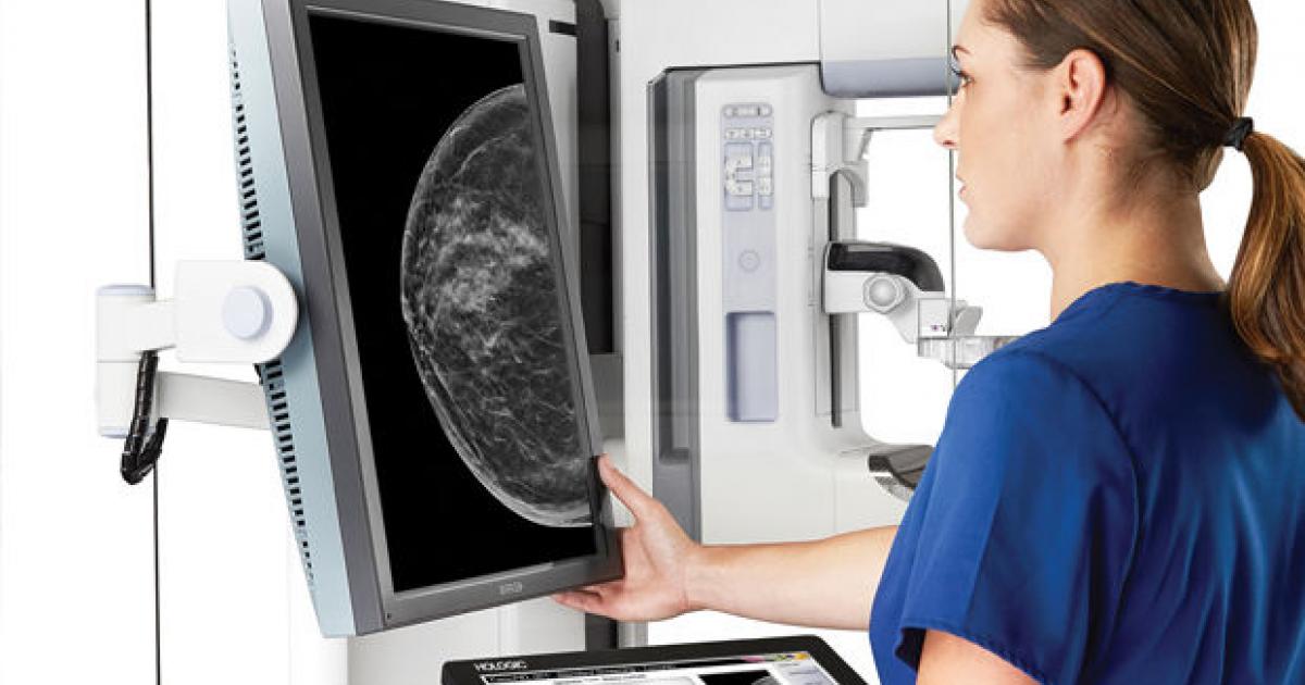
This is followed by an X-ray of the breasts, a mammographic examination. Mammography is an X-ray examination performed at low radiation exposure, which can be safely used in the case of repeated examinations thanks to direct digital technology. During the examination, the breasts are placed on the X-ray surface, the images are taken by using compression, from two angles, with low-energy ionizing radiation. The examination is painless, however, due to the compression of the breasts, depending on individual sensitivity, there may be some discomfort.
Tomosynthesis is a more modern method of examining the breast structure than mammography. During the examination, a millimetre-accurate image of the breast is obtained from the synthesis of layer images, therefore latent lesions can be detected more easily even in the case of dense breast tissue.
In case of palpable, circumscribed lesions, dens, complicated mammographic structures, or operated, implanted breasts, or in case of high-risk, breast ultrasound examination is also performed after mammography. For women under the age of 30, the examination is performed after the physical examination.
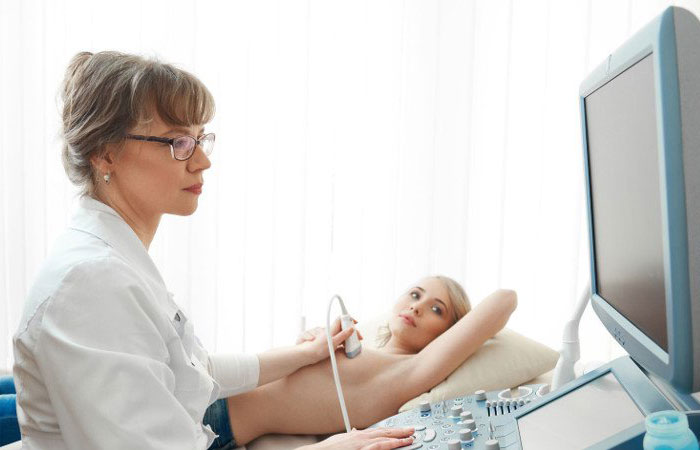
Breast ultrasound is completely painless: the radiologist uses a probe to direct the ultrasound waves to the tissues of the breast and axils, the reflected waves help get an accurate picture of the lesion.
It can be used to differentiate between tissue and cystic lesions, and to depict abnormal (inflamed or cancerous) milk ducts, lymphatic vessels, scars, and fluid deposits (haematoma, postoperative seroma, abscess).
The radiologist may also recommend sampling (biopsy) based on the findings. Sampling is performed in all cases, whether the lesion is palpable or not, using an imaging technique. If the lesion is clearly visible with ultrasound, ultrasound-guided sampling is recommended. Mammography-guided stereotaxic sampling is performed in the case of impalpable, ultrasound-indeterminate, or uncertain benign lesions.
The type of biopsy is determined in each case by the radiologist in the knowledge of the lesion found. Sampling methods can be puncture (aspiration of fluid) for cysts, cytological sampling (separation of cells by needle puncture), or core biopsy (excision of a tissue cylinder with a needle).
If none of the methods above provide a clear answer to all questions, a breast MRI scan can help clarify the diagnosis. Screening is only recommended for high-risk women. It is the primary examination method to rule out or prove damage to breast implants.
Findings from complex breast diagnostic examinations are evaluated by a radiologist.
