Joint MRI (knee, hip, shoulder, elbow)
The soft tissues surrounding the joints and bones can be perfectly examined wit an MRI scan.
Tendons, ligaments, cartilages, synovial membranes become visible. Joint MRI examination can be performed on the joints of the leg – knee, ankle –, arm – elbow, shoulder –, and hip.
For an accurate diagnosis, your doctor might request an MRI and an ultrasound examination as well.

What kind of preparation is required?
In all cases, it is recommended that you consult your doctor about the details before having an MRI scan. The native test requires no special preparation, but you should not eat for 6 hours before an examination with contrast agent!
It is important to know that due to the strong magnetic field, it is forbidden to bring any metal or device, object containing metal into the examination room! This also applies to jewellery and piercings in the body.
Are there any side effects of the examination?
MRI examination has no side effects, rarely the contrast agent can cause a reaction. In case of pre-existing known allergies, scan with a contrast agent is not performed.
Types of joint MRI examinations
-
Knee MRI
The examination of the bones that make up the knee, the articular cartilage, the cricoid cartilage of the knee joint (meniscus), the kneecap (patella), and the condition and position of the cartilage of the patella, and all the ligaments supporting the knee is covered by the knee MRI.
-
Hip MRI
It helps detect oedema, abrasions, degenerative processes, early femoral head necrosis, but the hip MRI also gives an image of the condition of the joint capsule.
-
Shoulder MRI
Abrasive diseases of the shoulder joint, injuries after accident, rupture of the rotator cuff surrounding the shoulder, articular surface, cartilage thickness, and soft tissue lesions can be well assessed with the shoulder MRI examination.
-
Elbow MRI
The doctor can see the state of the three bones meeting in the elbow, the lesions following the trauma, the inflammation of the bursa, the amount of fluid accumulating in the bursa, and diagnose the tennis elbow as a result of straining the extensor musle of the forearm.
The elbow MRI shows the tendons, cartilages, the condition of the periosteum, the inflammations and the changes they cause.
How is a joint MRI scan performed?
The MRI device resembles a large, thick ring with a table moving at the bottom, the patient is lying on this as instructed by the doctor.
Examination of the joints is usually done lying on your back. However, some tests may require tilting and retaining the joint to make the best shot.
Examination of the elbow joint is most often done with the inwardly tilted (pronated) forearm, holding the hand with the palm facing down. The MRI device is noisy, makes strange, knocking, clicking machine noises, but its intensity can be reduced by using earplugs.
Medicover’s MRI device is called a closed machine, so the patient may feel confined.
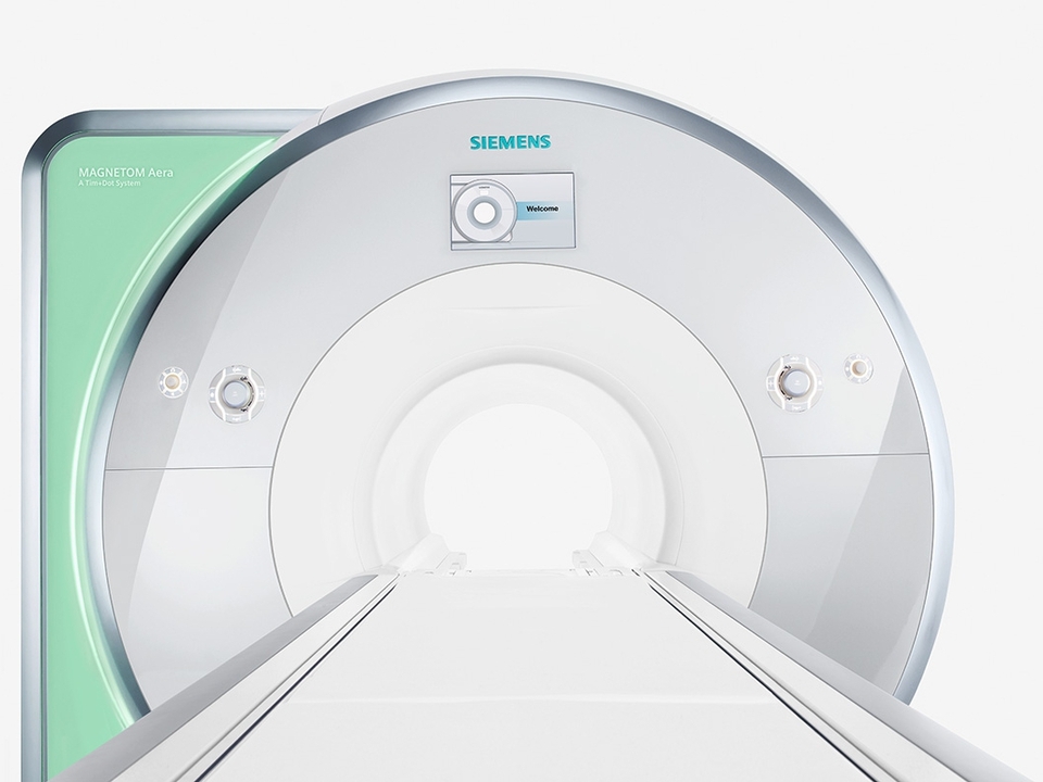
However, the good news is that this can be mitigated by the fact that the machine’s diameter is larger than average with its 70 cm. During the examination, the patient does not feel anything.
When can the test not be performed?
When any magnetically responsive materials (joint prosthesis, nail, screw, metal valve, pacemaker, tourniquet, implant, etc.) are in the body. These can move out of place during the examination in a strong magnetic field and, in severe cases, can cause injury, and the resulting image of the organ being examined can become invaluable. Nowadays, after surgeries, patients receive a document stating whether or not they can be examined with MRI with the built-in implant. We ask our dear patients to bring this document!
If the patient is prone to claustrophobia, the test may be difficult to be performed.
When is the result expected?
The completed scan will be reviewed by the radiologist and the result will be available in our online system after 5 working days.
