Thoracic spine MRI examination
MRI examination of the thoracic spine can be used to determine spinal lesions, disc diseases, bone marrow and intervertebral discrepancies, but it is also suitable for monitoring post-operative scarring.
In our institute, MRI examination of the thoracic section is most often used in case of suspicion of trauma or degeneration, or to detect processes affecting the spinal cord.
MRI is an imaging diagnostic procedure that uses magnetic field to take detailed images of the human body.
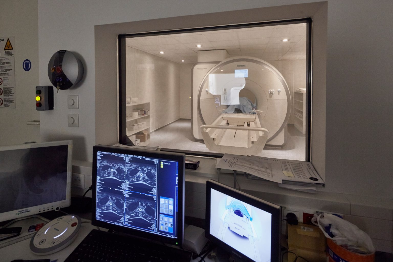
The magnetic field generated by the device converts the energy changes in the body into a high-resolution, three-dimensional image by a high-performance computer. At Medicover Diagnostic Center, we use equipment that works with a closed but extremely short tube, it is 10 centimetres wider than average, and its diameter is 70 cm, thus helping reduce the feeling of confinement.

What types of diseases does a spinal MRI scan help diagnose?
With an MRI scan, several types of diseases can be diagnosed. Your doctor may request an examination if you have any of the following complaints or illnesses.
- disc abrasion
- disc herniation
- cancerous disease
- inflammation
- injury
With which symptoms is it recommended to have the scan done?
Your doctor may consider an examination if you have long-term local pain and balance disorder, your gait is unsteady and you feel weakness in your lower extremities.
How to prepare for the thoracic spine MRI scan?
In all cases, it is recommended that the treating physician be consulted prior to the MRI examination, although native examination does not require special preparation.
It is important to know that due to the strong magnetic field, it is forbidden to bring any metal, device or object containing metal into the examination room!
This applies to jewellery, piercings in the body, and some makeup products may also contain substances that affects the success of the test.
Prior to an examination with contrast agent, specialist consultation is mandatory. Kidney function laboratory results showing creatinine and urea values are also required to perform this type of examination.
In the case of a scan with contrast agent, you should not eat for 6 hours beforehand.
How does the examination work?
Thoracic spine MRI is usually performed in supine position but can rarely be performed while the patient is lying on their abdomen or side, depending on their condition.
After the pre-examination orientation, the operator will accompany you into the examination room and then ask you to lie on the examination bed. The operator adjusts the correct body position, places the coils required for the examination, and then the examination bed lifts you into the magnetic tube. It is important to remain still during the scan, you can only change your body position on the instructions of the operator. The examination takes an average of 30 minutes, while you can communicate with our colleague continuously through the built-in microphone and speaker. The machine makes a loud, rattling sound that can be reduced with ear protection.
When is a thoracic spine MRI scan not possible to be performed?
- The examination cannot be performed in the first trimester of pregnancy.
- The examination cannot be performed if you do not have an MRI compatible metal, implant, prosthesis (pacemaker), orthopaedic metal (screw, plate, nail, wire), implanted joint prosthesis, prosthetic limb, implanted defibrillator, neurostimulator, implanted drug dispenser, vascular implants (e.g. clips, vascular occlusion devices), ear implants, metal foreign body (e.g. bullet, splinter, other electronic implants). Therefore, in case of an implant, implanted metal, prosthesis, please bring a written certificate of its compatibility with MRI examination (certified by the implanting specialist, institution) for the examination itself, which clearly states: the suitability to enter the MRI examination room, to perform the MRI examination and marking the maximum magnetic field strength (1.5 Tesla, 3 Tesla) for the device or implant. Inform the person performing the examination about the implant beforehand.
- If the subject weighs more than 200 kilograms, but it also depends on the circumference of the abdomen.
- In case of a scan with contrast agent, if the eGFR of the kidney function is below 45 ml/min, the test is not performed.

What to expect after the thoracic spine MRI scan?
The finding in itself is not a diagnosis. After the examination, please always consult a specialist with the result.
The contrast agent used for MRI examinations is gadolinium-based. In rare cases, hypersensitivity might happen, which may occur in the form of an allergic reaction immediately after administration of the contrast agent.
When is the result expected?
The completed scan will be reviewed by the radiologist and the result will be available in our online system after 3 working days.
